
Why Diagnostic Imaging Drives Accurate Vein Treatment Planning
Leg vein symptoms may appear simple at first, yet the cause is often hidden below the skin. Bulging veins, swelling, heaviness, cramps, and itching may reflect pressure changes inside deeper pathways. That is why accurate imaging matters before any treatment plan is chosen. A clear visual map helps vein specialists connect symptoms with blood flow patterns, measure disease severity, and select minimally invasive treatments that match each patient’s leg health needs.
Imaging Finds the Real Source
Many visible leg veins are a surface sign of a deeper circulation problem. Before treatment begins, specialists need to know which veins leak, which remain healthy, and how blood moves during rest and standing. A vein doctor in Hauppauge can use diagnostic imaging to study reflux, vein width, and valve function, giving each patient a plan based on evidence rather than guesswork alone.
Symptoms Can Mislead
Leg discomfort does not always match what appears on the skin. Small spider veins may cause cosmetic concern, while larger varicose veins can reflect chronic venous insufficiency. Some people also report aching, throbbing, or swelling without large visible veins. Imaging helps connect those complaints to actual changes in circulation. That step prevents assumptions and keeps treatment planning focused on the real source of leg symptoms.
Ultrasound Adds Measurable Data
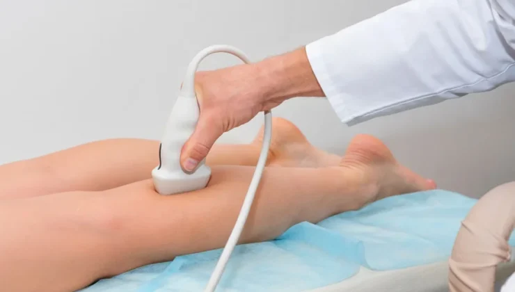
Duplex ultrasound gives specialists live information about blood flow and vein structure. It can show whether valves close well or allow backward movement, which raises pressure in the legs. Imaging also measures vein diameter and tracks the length of diseased segments. Those details matter because treatment choices depend on location, size, and severity. Better data support safer planning and clearer expectations.
Better Maps Improve Treatment Choice
Not all varicose veins begin in the same place. One patient may have reflux in a major surface vein, while another may have branch veins causing discomfort. Imaging creates a map that guides treatment choice with more precision. That can help determine whether radiofrequency ablation, sclerotherapy, adhesive closure, foam treatment, or microphlebectomy best fits the pattern found in the legs.
Timing Matters Too
Vein disease usually changes over time, and symptoms often worsen after long sitting, standing, pregnancy, or aging. Imaging helps show whether care should begin soon or whether supportive steps may be tried first. A specialist can compare symptom history with scan findings and decide the right sequence for care. That order matters because treating the main source first can improve comfort and results.
Planning Reduces Unneeded Care
Without imaging, treatment decisions may rely too heavily on appearance alone. That increases the chance of addressing visible veins while missing the source that feeds them. Diagnostic scans reduce that risk by showing the full pattern in the legs. A targeted plan can limit unnecessary treatment, improve efficiency, and support better symptom relief with fewer surprises during the care process.
Imaging Helps Rule Out Other Concerns
Leg swelling or pain may have more than one cause, and vein specialists need a clear picture before moving ahead. Diagnostic imaging can help identify signs of vein disease and help rule out certain urgent concerns, such as deep vein thrombosis, that require different medical management. That careful review protects patients from starting the wrong treatment path and keeps planning grounded in clinical facts.
Follow-Up Starts with a Strong Baseline
Good treatment planning does not begin on treatment day. It starts with a reliable baseline that shows how the legs function before care. Imaging provides that starting point and makes later comparisons more useful. Specialists can review symptom improvement against the original scan findings and clinical exam. Strong baseline data also help explain why some patients need one treatment while others benefit from a staged approach.
Insurance and Clinical Proof
Data from diagnostic imaging can support medical necessity when leg symptoms reflect venous disease rather than cosmetic concern alone. Findings such as reflux, enlarged veins, and valve failure give clinical proof that helps shape treatment planning and insurance review. That documentation also improves communication between patients and specialists. Everyone can see the reason behind the plan, which often leads to better understanding and stronger follow-through.
Conclusion
Diagnostic imaging is central to accurate vein treatment planning because it turns symptoms into measurable findings. For people with varicose veins, spider veins, swelling, or leg heaviness, that clarity can shape every next step. Imaging shows where the disease begins, how severe it is, and which minimally invasive treatments fit best. A thoughtful, evidence-based plan gives patients a better chance of lasting symptom relief and healthier legs.